Pregnancies are diagnosed in the 5th week most of the times. Because the missed period is the first sign of pregnancy in re-productively active females. Fetal age is calculated from the date of last menstrual period. It is approximately 3 weeks after conception. 5th week of pregnancy comes in the first trimester.
5th Week of Pregnancy Symptoms
- Amniotic fluid starts to build up
- Embryo gains the size of about 4-5 mm.
- The egg completely implanted in the uterine wall. The mucous membrane of uterus covers the egg, amniotic cavity and umbilical sac.
- Placental development continues
- A fibrin clot makes a plug in the hole of the uterine cavity
- The developing embryo starts receiving the nutrients and oxygen via the placenta
Why Ultrasound Scan at the 5th Week
5 week ultrasound what to expect – Ultrasound scan at 5th week is also known as dating scan. It is for calculating the expected date of delivery. Another method for estimation of the date of delivery is the date of last menstrual period. The other reasons for 3 weeks to 5 weeks ultrasound are:-
- For detection of abnormalities
- To confirm the pregnancy
- To monitor the growth of the fetus
- To rule out the twin pregnancy
- To locate the placenta because the low lying placenta may be problematic during normal labor. The mode of delivery might be cesarean due to low lying placenta i.e. placenta previa.
- To rule out ectopic pregnancy
The Procedure of 5 Week Ultrasound
You will be asked not to go to micturition before the scan. Because full bladder pushes the uterus upward, it gives a clearer picture. You will be asked to lie down on the table and a get will be applied to your abdomen. The doctor will then move a high frequency producing sound on your abdomen. The sound will be reflected from the uterus and creates a picture, which will be displayed on the screen.
What to expect at 5 Week Pregnant Ultrasound
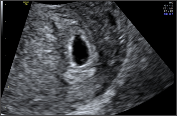
Fetal structures that can be identified at 5 weeks of pregnancy are embryo yolk sac and gestation sac. If you are having dizygotic twins the doctor will be able to identify the yolk sac and fetal poles present in two different sacs. But if you are having mono zygotic twin then there will be one gestational sac, two yolk sac, and embryos in the sac. By the end of week 5, the organogenesis has started. The major organs like stomach, heart, kidney, brain, and liver started their differentiation. This is the time when pregnant females should take care of drug or radiation exposure because it will directly affect the organs of the baby. The presence of the yolk sac is a confirmation of intrauterine pregnancy, and it rules out the ex-topic pregnancy. In ultrasound, the black area denotes the gestational sac and the white ring surrounding it denotes the yolk sac. The source of nutrition to the fetus is the yolk sac.
At the site of implantation, there are surrounding cavities known as the lacunary structure. Adjacent to the yolk sac is embryonic poles. It is because of the short connecting stalk. Sometimes the cardiac activity can be noted by trans-vaginal ultrasound. By the end of 5 weeks, the fetal heart rate is in the range of 60-90 beats per minute.
